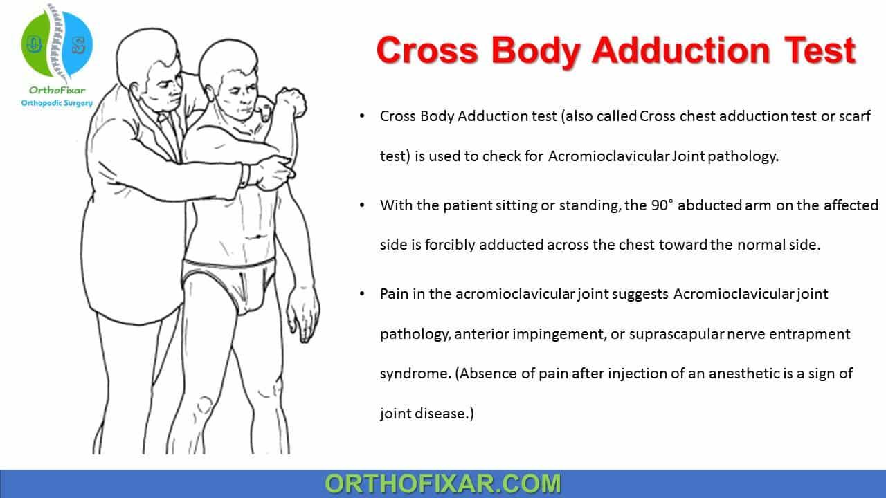
Cross Body Adduction test (also called Cross chest adduction test or scarf test) is used to check for Acromioclavicular Joint pathology.
In 1951, McLaughlin noted that many patients with AC joint pathologies developed a sharp pain at the top of the shoulder when the arm was actively adducted across the chest and towards the contralateral shoulder.
Bạn đang xem: Cross Body Adduction Test
How do you perform Cross Body Adduction Test?
With the patient sitting or standing, the 90° abducted arm on the affected side is forcibly adducted across the chest toward the normal side.
Several years later, Moseley described a modified version of this test in which the patient was asked to actively place the arm in a position of adduction as described by McLaughlin. The clinician would then apply an additional passive force to the patient’s elbow which effectively increased the amount of adduction and AC joint compression. Although this version of the test has not been validated by any study, it may be a useful adjunct to detect more subtle forms of AC joint-related pain when the diagnosis is unclear.
See Also: Clavicle Anatomy
What does a positive Cross Body Adduction Test mean?
Pain in the acromioclavicular joint suggests Acromioclavicular joint pathology, anterior impingement, or suprascapular nerve entrapment syndrome. (Absence of pain after injection of an anesthetic is a sign of joint disease.)
To confirm the diagnosis of AC joint-related pain, McLaughlin then injected the joint with local anesthetic, when repetition of the test following this injection was painless, it was determined that AC joint compression was causative and distal clavicle excision was recommended to alleviate the patient’s symptoms.
Dull, deep-seated pain over the superior scapular margin in the supraspinous fossa and posterolaterally on the scapula with radiation into the upper arm can be an indication of compression of the suprascapular nerve under the transverse scapular ligament by distal displacement of the scapula.
Sensitivity & Specificity
A study by Efstathios Chronopoulos for diagnostic value of physical tests for isolated chronic acromioclavicular lesions, he compared the three tests (acromioclavicular resisted extension test, active compression test and Cross Body Adduction Test) that is used in evaluation of acromioclavicular joint.
Xem thêm : Nearest major airport to Hawley, Pennsylvania:
The accuracy of Body Cross Test was as following:
- Sensitivity: 77 %
- Specificity: 79 %.
The study concludes that these tests have utility in evaluating patients with acromioclavicular joint pathologic lesions, and a combination of these physical tests is more helpful than isolated tests.
While there are many studies that have used the cross-body adduction test as a method of diagnosis, very few studies have been conducted with the purpose of evaluating the diagnostic utility of the cross body adduction test in patients with and without AC joint pain.
Maritz and Oosthuizen studied the test in a series of 22 patients with AC joint pain and calculated a sensitivity of 100 % using joint injection as the diagnostic standard. Chronopoulos et al. evaluated the clinical efficacy of the test in 35 patients who later underwent distal clavicle excisions. In that study, the sensitivity was 77 %, the specificity was 79 %, the positive predictive value (PPV) was 20 %, and the negative predictive value (NPV) was 98 %.
Another tests for Acromioclavicular Joint pathology include:
1. Acromioclavicular Injection Test:
Inject the acromioclavicular joint with an anesthetic such as lidocaine (with a corticosteroid where indicated) using a proximal approach.
The injection must be performed under sterile conditions. Large osteophytes, arthritic joints, or a defective meniscus may render the injection into the anatomically narrow acromioclavicular joint space impossible.
If the injection relieves local pain, at least temporarily, this indicates that acromioclavicular pathology is present. To confirm the diagnosis it is recommended, while anesthesia persists, to attempt to reproduce the pain with whichever examination produced the most pain prior to injection, such as the cross-body adduction stress test or painful arc test.
See Also: Subacromial Injection Test
2. Forced Adduction Test on Hanging Arm:
Xem thêm : Are Hot Tubs Bad for You? Here’s How To Soak Safely
The examiner grasps the upper arm of the affected side with one hand while the other hand rests on the contralateral shoulder and immobilizes the shoulder girdle. Then the examiner forcibly adducts the hanging affected arm behind the patient’s back against the patient’s resistance.
Pain across the anterior aspect of the shoulder suggests acromioclavicular joint disease or subacromial impingement. (Symptoms that disappear or improve following injection of an anesthetic indicate that the acromioclavicular joint is causing the pain.)
3. Clavicle Mobility Test:
The examiner grasps the lateral end of the clavicle between two fingers and moves it in all directions.
Increased mobility of the lateral clavicle with or without pain is a sign of instability in the acromioclavicular joint.
In isolated osteoarthritis there will be circumscribed tenderness to palpation and pain with motion.
Acromioclavicular joint separation with rupture of the coracoclavicular ligaments will be accompanied by a positive “piano key” sign: the subluxated lateral end of the clavicle displaces proximally with the pull of the cervical musculature and can be pressed inferiorly against elastic resistance.
Notes
Acromioclavicular (AC) joint is a plane/gliding joint with a fibrocartilaginous disc.
When the arm is maximally elevated, about 5 to 8 degrees of rotation is possible at the AC joint, although the clavicle rotates approximately 40 to 50 degrees.
The cross body adduction test is also used to measure the general shoulder motion and can be used more specifically to measure flexibility of the shoulder, especially with regard to the posterior capsule. Measuring tape can be used to measure the distance from the lateral elbow epicondyle to the AC joint at the top of the shoulder. Once this has been completed and a measurement has been recorded, the test is repeated on the contralateral side for measurement comparison.
Reference
- Efstathios Chronopoulos, Tae Kyun Kim, Hyung Bin Park, Diane Ashenbrenner, Edward G McFarland.Diagnostic value of physical tests for isolated chronic acromioclavicular lesions. Am J Sports Med . Apr-May 2004;32(3):655-61. doi: 10.1177/0363546503261723.PMID: 15090381
- McLaughlin HL. On the frozen shoulder. Bull Hosp Joint Dis. 1951;12(2):383-93.
- Moseley HF. Athletic injuries to the shoulder region. Am J Surg. 1959;98:401-22.
- Maritz NG, Oosthuizen PJ. Diagnostic criteria for acromioclavicular joint pathology. J Bone Joint Surg Br. 2002;78 Suppl 1:78.
- Chronopoulos E, Kim TK, Park HB, Ashenbrenner D, McFarland EG. Diagnostic value of physical tests for isolated chronic acromioclavicular lesions. Am J Sports Med. 2004;32(3):655-61.
- Clinical Tests for the Musculoskeletal System, Third Edition
- Millers Review of Orthopaedics, 7th Edition
Nguồn: https://buycookiesonline.eu
Danh mục: Info
This post was last modified on November 24, 2024 9:49 am

