Abstract
Introduction
The initiation of uterine contractions after embryo transfer that leads to the immediate or delayed expulsion of the embryos is considered one of the major factors that limit the success rates after IVF. It has been demonstrated that about 15% of the embryos are expelled after transfer ( Poindexter et al. , 1986 ) and about 42% of the injected dye in a mock embryo transfer is extruded ( Kuntzen et al. , 1992; , Mansour et al. , 1994 ).
An attempt was made by our team to prevent the expulsion of the embryos from the uterine cavity after embryo transfer by applying a gentle mechanical pressure on the portio-vaginalis of the cervix using the vaginal speculum ( Mansour, 2005 ). It is a simple modification of the embryo transfer technique, yet it significantly improves the implantation and clinical pregnancy rates. However, the study did not demonstrate why the higher pregnancy rate was obtained. Only a methodological approach looking at the outcome of mock embryo transfer demonstrating the displacement of markers following occlusion or no occlusion of the cervix might give an explanation. In the present study, we aimed to investigate further using ultrasonographically visible material to explain the mechanism of action of this technique. To the best of our knowledge, this is the first time in the medical literature that washed, concentrated spermatozoa have been used as an ultrasonically visible material.
Bạn đang xem: Sperm suspension is a highly ultrasonically visible material: a novel model to study uterine activity
Materials and Methods
Patients included in the study were infertile couples undergoing intrauterine insemination (IUI) for unexplained and mild male factor infertility at the Egyptian IVF-ET Center. Ethical approval for the study was obtained from the institutional review board. The patients were counselled that the study does not change any protocol or methodology of the IUI they were scheduled for. They were told that the IUI was going to be done under abdominal ultrasound guidance of the catheter and that there would be an extra 10 min before the IUI procedure to study uterine activity.
Procedure
All of the patients were subjected to transvaginal folliculometry to follow up the size of the leading follicle until it reached ≥ 18 mm. IUI was timed 34 h after hCG injection. The semen was prepared on the day of insemination using the swim-up technique as described previously ( Mansour et al. , 1989 ).
All patients were placed in the lithotomy position, no anaesthesia was used. After visualizing the portio-vaginalis of the cervix using Cusco’s speculum, the cervix was cleaned using sterile gauze. Insemination was done using a sterile Wallace catheter (1816 N, HG; Wallace Ltd, Colchester, UK) to mimic the embryo transfer procedure ( Mansour, 2005 ). The catheter was attached to a 1 ml tuberculin syringe and filled with 0.5 ml of a processed washed and concentrated sperm suspension.
In the study group ( n = 22), after introducing the catheter, the screw of the vaginal speculum was loosened so that its two blades would close gently on the portio-vaginalis of the cervix. After waiting for about 1-2 min, only 50 ul was injected into the uterine cavity. The catheter was then withdrawn slowly and the vaginal speculum was left in place pressing on the cervix after withdrawal of the catheter for 10 min before removal. The procedure was monitored for 10 min and video recorded for documentation and further evaluation using abdominal ultrasound (Medison 9900, Kretz, Austria).
Xem thêm : Bird Watcher’s General Store
In the control group ( n = 23), the same procedure as in the study group was followed, but no loosening of the screw of the vaginal speculum to close on the portio-vaginalis was done. The speculum was kept open and was removed after withdrawal of the catheter. Monitoring by ultrasound was performed for 10 min and video recorded for further evaluation.
In both groups, after completing the experiment and recording, the rest of the sperm suspension (0.5 ml) was then slowly injected as in a routine IUI. The patients were allowed to rest in bed for 15-30 min then go home.
The primary outcome measure was observing the location of the injected material in the upper uterine cavity and recording its passage to the lower uterine segment and through the cervical canal. For this small pilot study, findings were compared using Chi-square test and results are presented as Odds ratio with 95% confidence interval (CI).
Results
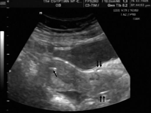
The sperm suspension was clearly visible in all cases. In the closed speculum group, the echogenic injected droplet remained in the upper uterine segment in 18 cases (82%) and moved towards the lower uterine segment in six cases (18%) (Fig. 1 ). In the open speculum group, the echogenic droplet remained in the upper uterine segment in only six cases (26%) and it moved towards the lower uterine segment (Fig. 2 ) and passed through the cervical canal in 17 cases (74%) [OR = 12.75, 95%, CI = 3.1-53.2 ( P < 0.0003)].
Fine peristaltic movements were observed moving the ultrasonically visible droplets. In the closed speculum group, the direction of the movements was from the lower to the upper uterine segment. In two cases the echogenic droplets moved towards the lower uterine segments, then it was pushed again upwards towards the fundus. In the open speculum group most of the movements of the echogenic droplets were towards the lower segment and the cervix.
The whole amount of sperm suspension was then injected for IUI, and Fig. 3 shows the echogenic droplet in the fundus and Fig. 4 shows the echogenic sperm suspension filling the whole cavity.
Discussion
Xem thêm : Purina Moist and Meaty Dog Food Review (Semi-Moist)
The immediate or delayed expulsion of embryos after transferring them into the uterine cavity has always been a major concern in assisted reproduction ( Harper et al. , 1961 ; Menezo et al. , 1985 ; Poindexter et al. , 1986 ; Schulman, 1986 ; Meldrum et al. , 1987 ). In two different studies mimicking embryo transfer, one using a radio opaque dye ( Knutzen et al. , 1992 ) and the other using a methylene blue dye ( Mansour et al. , 1994 ), it was found that the dye was extruded from the uterine cavity in 42% of the cases. Using artificial dyed embryos for training in IVF, it was also found that only 45% of the embryos were present within the uterine cavity 1 h after the transfer ( Menezo et al. , 1985 ). It has also been observed that after embryo transfer, the embryos can just as easily move towards the cervical canal as towards the Fallopian tubes ( Woolcott and Stanger, 1997 , 1998).
These findings from previous studies have raised concerns about the possibility of losing embryos after the embryo transfer, and inspired us to develop a technique to reduce embryo expulsion after embryo transfer by applying gentle pressure on the portio-vaginalis of the cervix using the vaginal speculum ( Mansour, 2005 ). However, our previous study did not provide a mechanism by which the higher pregnancy rate was obtained using this modified technique. Only a methodological approach looking at the outcome of mock embryo transfer demonstrating displacement of the markers following the occlusion or no occlusion of the cervix might provide an explanation. The improvement in the results in our previous study ( Mansour, 2005 ) may have resulted from phenomena not directly linked to attempting to occlude the cervix, and therefore the present study was performed. The results of this new study indicate that the gentle mechanical closing of the portio-vaginalis of the cervix using the vaginal speculum prevents extrusion of the injected material inside the uterine cavity to the cervical canal. In addition, this is the first study in the medical literature that demonstrates the echogenecity of washed, concentrated sperm suspension and its possible detection under ultrasound guidance. This finding by itself will be of value for using it for any further experimental study to evaluate uterine contractions. It is a very simple idea because concentrated sperm suspension is highly echogenic as well as safe and economic and any study can be done during IUI.
In semen processing for IUI, the sample is washed twice with tissue culture media. After centrifugation, the supernatant is discarded, while the pellet is resuspended in tissue culture media. This technique is suggested to remove the seminal plasma and its various contents of prostaglandins. The prostaglandins content of the washed sperm suspension had an insignificant effect on contractility ( Sinnemaa et al. , 2005 ).
In our search for an ultrasonographically visible material to be injected inside the uterine cavity, it was found that Lesny et al. (1998) used echovist material. We tried to obtain it but it is not available in Egypt. It was also found that de Ziegler et al. (2003) used an ultrasonically visible material to study the uterine activity but unfortunately the composition of the material was not mentioned.
The mechanical theory of occluding the cervix is supported by other investigators who used the speculum to prevent fluid regurgitation from the cervix in Fallopian sperm perfusion ( Mamas, 1996 ). The idea of using the vaginal speculum to press on the portio-vaginalis of the cervix originally came from our observation during IUI. We have always observed that some of the injected sperm suspension regurgitated from the cervix after injection and we modified our IUI technique by loosening the screw of the vaginal speculum after introducing the IUI catheter and before injecting the sperm suspension. When the valves of the speculum closed on the portio-vaginalis, the injected fluid did not escape through the cervix. The change in the uterocervical angle due to the release of the speculum may be another added factor for explaining this improvement.
The presence of subendometrial peristaltic movement was described by Birnholz et al. , 1984) . Endometrial wave-like activity was observed in 71% of spontaneous cycles and 91% of stimulated cycles and their direction from cervix to fundus usually benefits implantation ( Ijland et al. , 1998 , 1999 ). In our study group (closed speculum), the echogenic droplets moved in a wave-like movement in the direction from cervix to fundus.
In conclusion, this is the first time in the medical literature that concentrated sperm suspension has been used as a highly echogenic material that can be detected with ultrasound. The study provided evidence that pressing the portio-vaginalis of the cervix with the two blades of the vaginal speculum prevents the extrusion of injected material in the uterine cavity.
References
Nguồn: https://buycookiesonline.eu
Danh mục: Info
This post was last modified on December 2, 2024 3:46 pm

